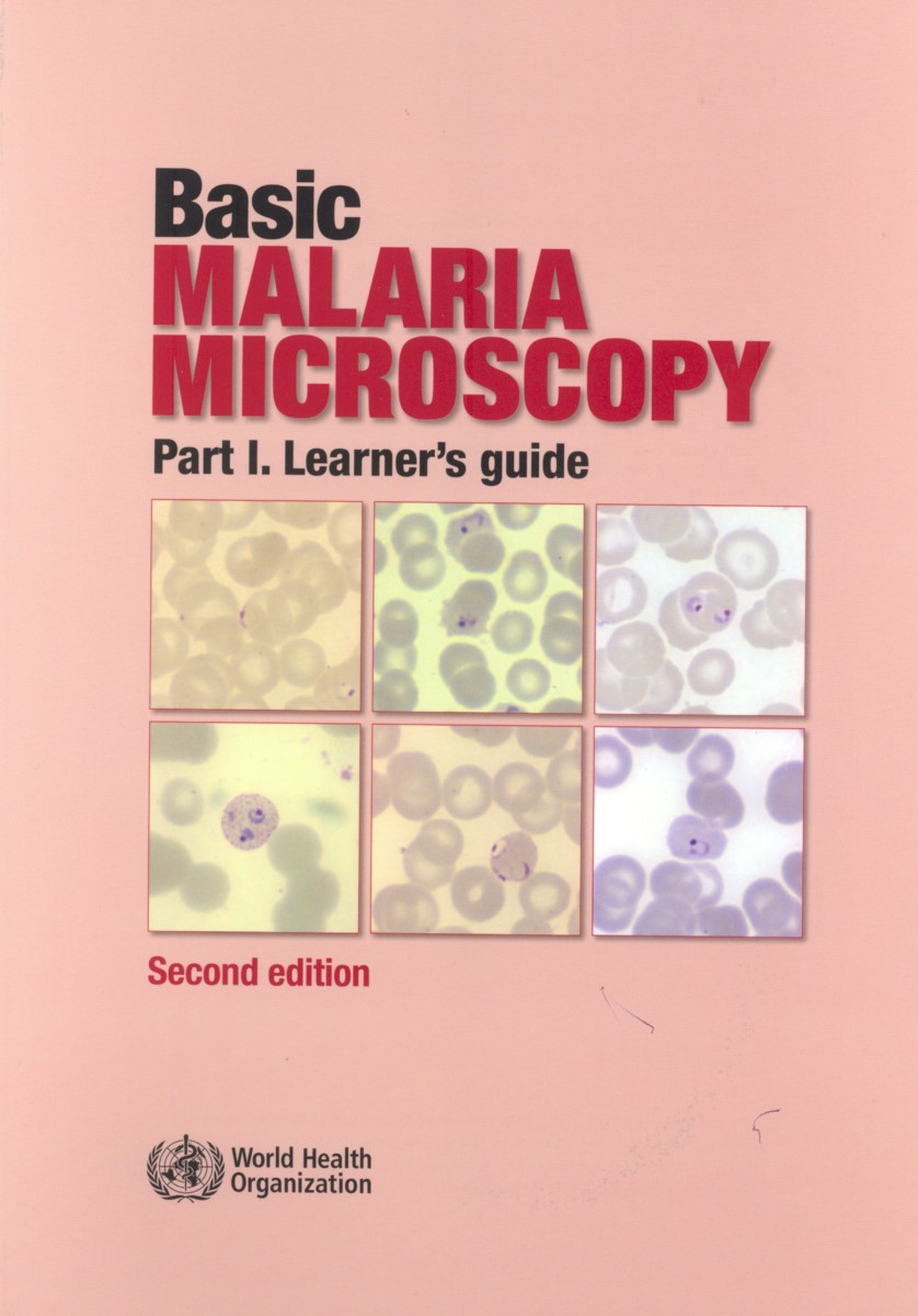
Basic Malaria Microscopy Edition 2
Part I. Learner's Guide
- Publisher
World Health Organization - Published
6th April 2010 - ISBN 9789241547826
- Language English
- Pages 83 pp.
- Size 8.25" x 11.75"
- Images color illus
Microscopists are vital to malaria programs, and their diagnostic and technical skills are relied on in both curative services and disease surveillance. Thus, training in malaria microscopy must be sound and must reach today's high standards.
This training package has been adjusted to meet the changes in the way malaria is diagnosed and treated. The training manual is divided in two parts: a learner's guide (Part I) and a tutor's guide (Part II). The package includes a CD-ROM, prepared by the United States Centers for Disease Control and Prevention, which contains microphotographs of the different malaria parasite species and technical information in PowerPoint format, which can be shown during training sessions and referred to by the participants. Emphasis is placed on teaching and learning, including monitoring and evaluating individuals and the group during training.
This handbook (Part I of the Basic Malaria Microscopy training modules) will assist participants during training in the microscopic diagnosis of human malaria. Designed as the foundation for formal training of 4-5 weeks duration, the guide is destined for participants with only elementary knowledge of science.
This second edition of the Basic Malaria Microscopy package is a stand-alone product, providing all that is needed to conduct a complete training course. It still contains the beautiful and accurate water-color illustrations prepared for the first edition of the manual by the late Yap Loy Fong. Experience has shown that color drawings are best in training new recruits to recognize parasite stages and species, because single plane pictures help students to extrapolate from what they see under the microscope, focused at a number of focal planes, to a complete view of the parasite. Later, they can move from drawings and use microphotographs, which will have an additional, positive impact. Thus, the training course is further strengthened if copies of the WHO Bench aids for malaria microscopy are also made available to trainees.
Link to Tutor's guide: Basic Malaria Microscopy: Part II. Tutor's guide
Preface
Introduction
1) Malaria, the Disease
2) Cleaning and Storing Microscope Slides
3) Keeping Records
4) Preparing Blood Films
5) Staining with Giemsa Stain
6) The Microscope
7) Examining Blood Films
8) Examining Blood Films for Malaria Parasites
9) Routine Slide Examination
10) Supervision in Malaria Microscopy
World Health Organization
World Health Organization is a Specialized Agency of the United Nations, charged to act as the world's directing and coordinating authority on questions of human health. It is responsible for providing leadership on global health matters, shaping the health research agenda, setting norms and standards, articulating evidence-based policy options, providing technical support to countries, and monitoring and assessing health trends.


